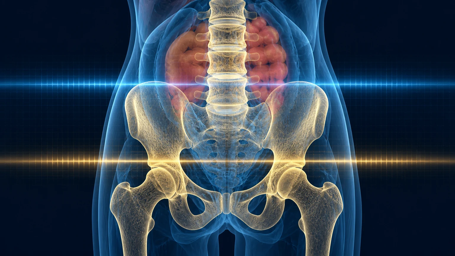
A DEXA scan may measure bone mineral density or estimate body composition. Learn how to read T-scores, Z-scores, lean mass and visceral fat without turning every decimal into a diagnosis.
You receive a report full of decimals: T-score, Z-score, lean mass, body-fat percentage and perhaps a visceral-fat estimate. The immediate question is: “is this good or bad?” The difficulty is that a DEXA scan does not answer one question, and its numbers do not all mean the same thing.
It may be a hip-and-spine examination for bone, or a whole-body scan for estimated body composition. They share technology but not purpose. A useful reading begins before you lie on the table: are you estimating fracture risk, monitoring lean-mass loss or following a body-composition intervention?
What matters before reading the report
- DXA is the technical acronym; DEXA is the better-known form. They refer to the same technology.
- A T-score describes bone density, not “bone age”.
- Lean mass is not the same as muscle and cannot replace a strength test.
- Comparing two scans requires the same scanner, protocol and the facility's measurement error.
Two tests under one familiar name
DXA uses two X-ray energies. For bone, it calculates areal mineral density in g/cm². In a whole-body scan, software partitions the image into bone mineral, fat and lean tissue. Results rely on manufacturer models and region placement, so changing scanner or software may break the comparison.
| Result | What it represents | What it cannot prove alone |
|---|---|---|
| BMD | Projected bone mineral per area at the hip, spine or forearm | Complete bone quality or total personal fracture risk |
| T-score / Z-score | Comparison with a reference population | Biological age or a treatment decision by itself |
| Lean mass | Tissue that is neither fat nor mineral | Pure contractile muscle, strength or power |
| Total and regional fat | Estimated mass and percentage by region | Inflammation, liver fat or individual cardiometabolic risk |
| Visceral fat | Algorithmic estimate of internal abdominal fat | Direct anatomical segmentation such as MRI |
Bone: how to read a T-score and Z-score
In postmenopausal women and men aged 50 or older, the T-score is preferred. ISCD states that a value of -2.5 or lower at the femoral neck, total hip or lumbar spine can establish densitometric osteoporosis. The 33% radius is used in selected circumstances. A score between -1 and -2.5 describes low bone mass, but that label alone does not decide whether treatment is needed.
Before menopause and in men younger than 50, the Z-score is preferred. A value of -2.0 or lower is described as “below the expected range for age”. In men younger than 50, bone density alone cannot diagnose osteoporosis.
The number belongs in a wider map. NIAMS summarises that, in studied populations, fracture risk rises about 1.5 to 2 times for each one-point fall in T-score. That is a relative change, not a personal probability. Age, falls, previous fractures, corticosteroids, low weight, menopause and other conditions may matter as much as the result.
Who may benefit from a bone density scan?
Recommendations vary by country and health system. The 2025 USPSTF recommends screening women aged 65 or older and younger postmenopausal women at increased risk after clinical assessment. For men, it finds population-screening evidence insufficient. Local guidance and personal history still determine the decision.
A DXA scan may be particularly useful with:
- a low-trauma fracture, height loss or suspected vertebral fracture;
- early or surgical menopause plus additional risk factors;
- treatments that accelerate bone loss, such as corticosteroids in selected contexts;
- low weight, malabsorption, bariatric surgery, hypogonadism, immobilisation or a condition associated with bone loss;
- starting or monitoring osteoporosis treatment when the result can change management.
The scan does not replace asking about falls, nutrition, exercise, medication or fractures. The person's risk is treated, not an isolated image.
Body composition: lean mass is not the same as muscle
Whole-body DXA can add lean tissue in the arms and legs as appendicular lean mass. That measure helps confirm low muscle quantity, but sarcopenia is first suspected from low strength. The EWGSOP2 consensus uses muscle quantity to confirm the diagnosis and physical performance to grade severity.
The distinction is practical. “Lean mass” includes water, skin, organs and connective tissue. In a 2026 analysis of 32,961 UK Biobank participants, DXA estimated fat reasonably but overestimated lean tissue compared with MRI. Among 1,928 people rescanned about 2.5 years later, MRI detected a 4% to 5% decline in muscle and lean mass that DXA did not capture.
During substantial weight loss, a report saying that lean mass is “maintained” is not enough to assume muscle is protected. Training, food intake, symptoms and functional strength deserve tracking too.
Visceral fat: useful for trends, not a verdict
Some scanners estimate visceral adipose tissue within the abdominal region. In 4,558 UK Biobank participants, that estimate correlated strongly with three-dimensional MRI volume, with an R² of 0.94. Performance was weaker in people with a BMI below 20. Strong population correlation does not make the two methods interchangeable for every individual.
A sensible reading uses visceral fat as one layer alongside waist circumference, blood pressure, glucose, triglycerides and ApoB. There is no universal DEXA threshold that independently diagnoses cardiometabolic risk. For trends, the same scanner and protocol help.
DEXA, bioimpedance, CT, MRI or function tests?
| Method | Strength | Main limit | Best fit |
|---|---|---|---|
| DEXA | Bone and regional composition at very low dose | Two-dimensional model; lean mass and visceral fat are estimates | Bone question or standardised composition follow-up |
| Bioimpedance | Fast, accessible and radiation-free | Strongly dependent on equation, hydration and device | Frequent tracking under the same conditions |
| CT | Detailed muscle, fat and infiltration | More radiation | An already indicated scan or specialist case |
| MRI | Three-dimensional tissue volume and quality without radiation | Cost, time and access | Research or a complex clinical question |
| Function | Measures what a person can do | Protocol and effort dependent | Whenever muscle or independence is the goal |
How to prepare for a useful comparison
Bone densitometry is quick, non-invasive and needs little preparation. RadiologyInfo advises telling the centre if pregnancy is possible or if you recently had barium, contrast material or a radiopharmaceutical. The centre may ask you to remove metal and pause calcium supplements for a specific period, so follow its instructions.
For comparable body-composition scans, try to reproduce:
- the same scanner, software and, if possible, operator;
- a similar time and similar food and hydration conditions;
- recent exercise, clothing and bladder status;
- positioning and analysed regions.
ISCD says each facility should calculate its precision error and least significant change, or LSC. If the difference between two tests does not exceed that threshold, it may be noise. A reported 300-gram change does not always mean 300 grams of real muscle or fat.
How often is a repeat worthwhile?
There is no universal “longevity” schedule. For bone, timing depends on the baseline result, treatment, fractures and expected rate of change. Intervals may shorten when rapid loss is plausible; in stable conditions, repeating too soon mostly measures noise.
For body composition, a baseline and another scan after an intervention long enough to matter may help if the answer can alter the plan. Scanning every few weeks rarely adds something that strength, waist, weight, symptoms and adherence could not show earlier.
| Question | Reasonable first measure | What to add |
|---|---|---|
| Is fracture risk the concern? | Central hip-and-spine DXA | Fractures, falls, secondary causes and clinical risk |
| Is there weakness or suspected sarcopenia? | Strength and performance first | DXA or bioimpedance for muscle quantity |
| Is substantial weight loss expected? | Baseline composition if it changes the plan | Strength, protein, symptoms and rate of loss |
| Is abdominal fat the concern? | Waist and clinical markers | DEXA as an added layer, not a diagnosis alone |
| Is it curiosity with no planned decision? | A scan is probably unnecessary | Define what result would change behaviour first |
Risks and signs that should not wait for DEXA
Direct harm is small: the test is painless and uses very little radiation. More common risks are indirect, including false reassurance, anxiety over decimals, artefacts from degeneration or prostheses, incompatible scanner comparisons and repeat testing that changes no decision.
A fall followed by hip pain and inability to bear weight, severe sudden spinal pain, rapid height loss, progressive weakness, unintentional weight loss or repeated fractures needs clinical assessment. A scheduled DEXA does not replace radiographs, blood tests or urgent care.
Frequently asked questions about DEXA scans
Are DEXA and bone density scans the same thing?
They use the same technology, but may be different protocols. Central hip-and-spine DXA measures bone mineral density; whole-body DXA estimates bone mineral, lean tissue and fat by region. Check which study the request includes.
What is the difference between a T-score and a Z-score?
A T-score compares bone density with a young-adult reference and is used mainly in postmenopausal women and men aged 50 or older. A Z-score compares with people of similar age and sex and is generally preferred before those groups.
What T-score means osteoporosis?
In postmenopausal women and men aged 50 or older, a T-score of -2.5 or lower at a valid site can establish densitometric osteoporosis. Fragility fractures and clinical risk also matter, even when the score is higher.
Does DEXA measure actual muscle?
It measures lean tissue, which includes muscle but also water, skin, organs and connective tissue. Muscle health also requires strength and performance. An image alone cannot tell how well you rise, carry or walk.
How reliable is DEXA for visceral fat?
Its estimate correlates well with MRI in population studies, but depends on the algorithm, scanner and body being measured. It is better for trends under the same protocol than as a stand-alone cardiometabolic diagnosis.
How much radiation does a DEXA scan use?
It uses a very small dose of ionising radiation and is quick and non-invasive. Exposure is not zero. Tell the centre if you are or may be pregnant and if you recently received contrast material or a radiopharmaceutical.
Do you need to fast before a DEXA scan?
Fasting is usually unnecessary for bone density, although the centre may give specific instructions. For comparable body-composition results, reproduce timing, hydration, food, exercise, clothing and bladder status.
How often should you repeat a DEXA scan?
There is no universal interval. It depends on the initial result, fractures, treatment and expected rate of change. A repeat is worthwhile only when change may exceed scanner error and alter a decision.
References
- International Society for Clinical Densitometry. Official Adult Positions 2023.
- Slart RHJA, Williams M, Teh J, et al. “Updated practice guideline for dual-energy X-ray absorptiometry.” European Journal of Nuclear Medicine and Molecular Imaging. 2025;52:539-563. Full text.
- U.S. Preventive Services Task Force. Osteoporosis to Prevent Fractures: Screening. 2025.
- National Institute of Arthritis and Musculoskeletal and Skin Diseases. Bone Mineral Density Tests: What the Numbers Mean. Accessed 29 August 2026.
- Radiological Society of North America and American College of Radiology. Bone Density Scan (DEXA or DXA). Reviewed 2024.
- Cruz-Jentoft AJ, Bahat G, Bauer J, et al. “Sarcopenia: revised European consensus on definition and diagnosis.” Age and Ageing. 2019;48(1):16-31. Full text.
- Zhang Y, Chen B, Gao Y, et al. “Muscle Imaging Assessments in Sarcopenia.” Calcified Tissue International. 2025. Full text.
- Paris M, Ifrim M, Lungren MP, et al. “Comparing DXA and MRI body composition measurements in cross-sectional and longitudinal cohorts.” Communications Medicine. 2026. Full text.
- Chowdhury A, et al. “Towards visceral fat estimation at population scale.” Frontiers in Endocrinology. 2023;14:1211696. Full text.
This article is educational. The indication, interpretation and timing of a DEXA scan depend on age, history, treatment, fracture risk and the clinical question. Pregnancy or its possibility should always be reported to the centre.
A good measurement does not give you a grade. It helps you make one specific decision and, when repeated, distinguish real change from noise.
Related articles

What Semen Can Tell You About a Man's Health
A semen analysis may open questions about testicular health, hormones, medicines and exposures. It is neither a fertility verdict nor a longevity test.
Can a Multivitamin Slow Biological Age? What COSMOS Measured—and What It Did Not
COSMOS found a small shift in two of five epigenetic clocks after two years. Here is the effect size, the limits and when supplementation answers a real nutritional need.

Cardiac Rehabilitation: Much More Than Exercise After a Heart Attack
Cardiac rehabilitation combines prescribed exercise, medication review, nutrition, psychological support and risk-factor control. Learn when to start, how long it lasts and how it changes with age or frailty.
uACR and eGFR: Two Tests That Reveal Hidden Kidney and Cardiovascular Risk
A normal creatinine does not rule out albuminuria. Learn how to read uACR and eGFR together, when to repeat them, and what a persistent result may change.